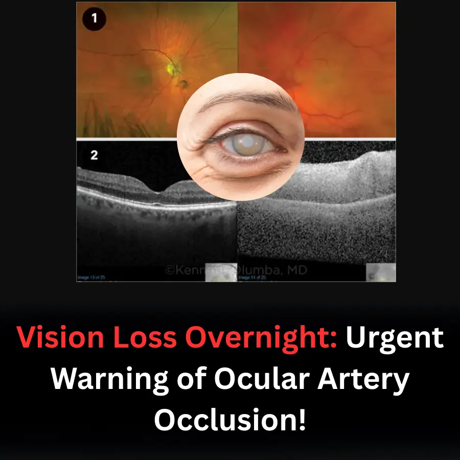
Waking up from a peaceful night’s sleep only to find complete darkness — total loss of vision — was the terrifying experience of 79-year-old Donald Dunes. His case illustrates that ophthalmic artery occlusion (OAO) is a true ophthalmic emergency, closely linked with cardiovascular disease, and requires rapid recognition and intervention within a narrow “golden window” of just a few hours.
An unusual event in the middle of the night
After watching late-night news, Mr. Dunes, an American man with a history of hypertension and dyslipidemia, went to bed as usual. In the middle of the night, he suddenly awoke with complete, painless vision loss in his left eye. His right eye remained entirely normal.
Assuming it was merely a bad dream, he tried to sleep again, but his condition did not improve by morning. Two days passed without any visual recovery before he finally called emergency services.
Emergency evaluation and initial findings
At the local hospital, a CT scan of the brain revealed no hemorrhage or major infarction. CT angiography of the brain and neck detected 80–90% stenosis of the left internal carotid artery. His ECG appeared normal. He was transferred to a tertiary emergency unit and examined immediately by an ophthalmologist.
Examination revealed:
-
Right eye vision: 20/20; Left eye: no light perception.
-
RAPD 3+ in the left eye.
-
Intraocular pressure: 12 mmHg (right), 9 mmHg (left).
-
Mild conjunctival inflammation and edema in the left eye with no iris neovascularization.
-
Fundus of the left eye: pale optic disc, diffusely white retina, narrowed retinal arterioles, and absence of a cherry-red spot.
OCT imaging showed a normal right eye but diffuse retinal edema and acute middle maculopathy in the left eye. Due to low intraocular pressure, emergency ocular massage was performed in an attempt to dislodge the embolus. However, because the patient arrived several days after symptom onset, thrombolytic therapy with Alteplase was no longer an option.
Additional imaging and laboratory findings
-
Brain and orbital MRI demonstrated multiple ischemic lesions in the left hemisphere, including frontal, parietal, occipital, caudate, and basal ganglia regions.
-
Echocardiography showed mild concentric hypertrophy, an ejection fraction of 60–65%, and otherwise normal cardiac function.
Differential diagnosis and final conclusion
Given the painless, sudden visual loss, the medical team considered several ischemic causes:
-
Central retinal artery occlusion (CRAO)
-
Ocular ischemic syndrome due to carotid stenosis
However, the features of diffuse retinal whitening, absence of a cherry-red spot, and severe ischemia affecting both the retina and choroid were more consistent with ophthalmic artery occlusion (OAO).
Because the patient was over 50, giant cell arteritis was ruled out with ESR, CRP, and CBC — all normal.
Final diagnosis: Left ophthalmic artery occlusion caused by severe stenosis of the left internal carotid artery.
Treatment and follow-up
Mr. Dunes received dual antiplatelet therapy (aspirin + clopidogrel) and underwent carotid endarterectomy ten days after diagnosis. Three months later, his left-eye vision remained unchanged. He continued to be monitored closely for complications such as iris neovascularization, anterior chamber changes, and retinal neovascularization — common sequelae of retinal ischemia.
Professional discussion
Ophthalmic artery occlusion is a vision-threatening emergency that occurs when blood flow to both the retina and choroid becomes blocked, most commonly due to carotid artery disease or cardiac emboli such as clots from atrial fibrillation.
Pathophysiology:
The ophthalmic artery arises as the first branch of the internal carotid artery, making it vulnerable to emboli, cholesterol plaques, or microthrombi.
Common risk factors:
-
Hypertension
-
Diabetes
-
Dyslipidemia
Clinical presentation:
-
Sudden, painless vision loss — ranging from severe impairment to complete blindness within minutes.
-
Fundus findings include diffuse retinal whitening and widespread ischemia without a cherry-red spot, distinguishing it from CRAO.
Treatment:
No therapy has been definitively proven to reverse vision loss after the acute phase. However, if the patient presents within 4–5 hours, IV Alteplase may improve vision (44% improvement vs. 13% without treatment).
Other interventions such as ocular massage, lowering intraocular pressure, or anterior chamber paracentesis remain supportive with limited evidence. Hyperbaric oxygen therapy may help if initiated within 6–8 hours.
Key clinical lessons
-
Ophthalmic artery occlusion should be treated as both an ocular and neurological stroke.
-
Time is critical — every minute of ischemia results in irreversible loss of retinal neurons.
-
After acute management, cardiovascular risk factors must be aggressively controlled to prevent recurrence.
-
Complications of retinal ischemia, including neovascularization and neovascular glaucoma, must be monitored and treated with anti-VEGF injections and panretinal photocoagulation when indicated.