A 16-Year-Old Girl Was Rushed to Hospital After a Pen Accident—What Doctors Discovered Inside Her Pelvis Shocked Them
A seemingly ordinary accident turned into an emergency operation after doctors discovered that a pen had traveled deep into a teenager's pelvic region.
It began with an accident that nobody could have imagined would lead to an emergency operation.
A 16-year-old student was reportedly at home in Bouaké, Côte d'Ivoire, when she fell while holding a ballpoint pen. What happened next would leave her experiencing severe pain and lead her family to seek urgent medical assistance.
At first, the situation may have appeared like an unusual household accident. But the consequences were far more serious than anyone initially expected.
The teenager had suffered an injury involving a foreign object that had entered through the vaginal area. Attempts to remove it did not resolve the problem. Instead, the object moved farther into her body, making the situation increasingly complicated.
When she eventually arrived at the hospital, doctors faced a medical challenge that required more than an ordinary examination.
Imaging tests revealed that the pen had traveled into the pelvis, close to important internal structures. The object was no longer accessible through a simple external examination, and doctors needed to determine its precise location before deciding how to remove it safely.
What followed was an emergency surgical procedure.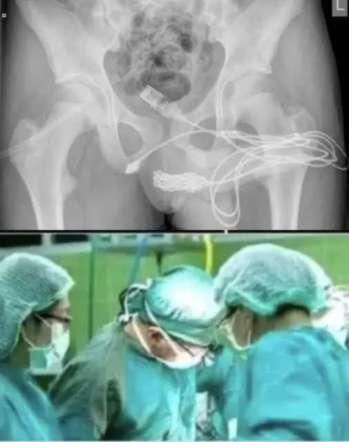
The case, documented by medical specialists at the University Hospital Center of Bouaké, highlights an unusual but important type of injury involving foreign objects and the female reproductive system.
But there is another detail that makes this story particularly concerning: the object was not simply lodged in one place. Its unusual path brought it close to the bladder and caused additional internal injuries.
How could an ordinary ballpoint pen create such a serious medical emergency? Why did the initial attempts to remove it make the situation more complicated? And what did doctors discover when they finally examined the teenager through medical imaging?
The answers reveal why even an apparently small accident involving a foreign object should never be treated casually.
An Ordinary Accident That Quickly Became a Medical Emergency
According to the published medical report, the patient was a 16-year-old student living in Bouaké, a city in central Côte d'Ivoire.
The incident reportedly occurred at home.
She was holding a ballpoint pen when she fell from standing height. During the fall, the pen accidentally entered her vaginal area.
This distinction is important: the medical publication describes an accidental injury, not an intentional attempt to insert an object.
Initially, the teenager experienced significant pain and vaginal bleeding.
These symptoms were concerning enough to prompt medical attention.
However, the situation became more complicated when attempts were made to remove the object.
The report describes approximately ten attempts by the teenager and medical personnel at an initial maternity facility to retrieve the pen.
Unfortunately, those attempts did not succeed.
Instead, the object disappeared farther into the body, making it much more difficult to locate and remove.
This is an important lesson in emergency care.
When an object becomes lodged inside the body, repeatedly attempting to pull it out without understanding its position can create additional injury.
The object may move deeper, damage surrounding tissue, or become positioned near organs that require special protection.
In this teenager's case, the medical team eventually needed to determine exactly where the pen had traveled.
Why the First Examination Was Not Enough
When the patient arrived at the University Hospital Center of Bouaké, doctors performed a clinical assessment.
The medical report noted that she was hemodynamically stable, meaning her circulation was not showing signs of immediate cardiovascular collapse at the time of examination.
However, that did not mean the injury was minor.
Doctors identified an important finding during examination: an end of the ballpoint pen could be felt through the suprapubic region, the lower abdominal area above the pubic bone.
Another significant problem involved the urinary bladder.
The teenager had developed urinary retention, meaning she was unable to empty her bladder normally.
Medical staff inserted a urinary catheter, allowing approximately two liters of urine to be drained.
This was a substantial amount and demonstrated how seriously the injury was affecting normal urinary function.
The examination also failed to reveal an obvious entry opening or active bleeding during the speculum examination.
These findings made medical imaging especially important.
Doctors needed more information before deciding how to proceed.
The object could not simply be pulled out based on what was visible during the initial examination.
Its position, direction, and relationship to surrounding organs had to be established first.
And this is where the X-ray and other imaging examinations became crucial.
The X-Ray That Revealed the Hidden Problem
The most important turning point in the documented case came when doctors performed imaging examinations.
A plain abdominal radiograph showed an unusual tubular structure in the right side of the pelvis.
The appearance suggested that an elongated foreign object was present.
Further examinations, including ultrasound and computed tomography, helped establish its location.
The CT scan provided additional information about the object's relationship to nearby structures.
For doctors, this was essential.
The pelvis contains several closely positioned organs and anatomical structures, including the urinary bladder, reproductive organs, blood vessels, and portions of the digestive system.
An object traveling through this area can create complications even when the external injury appears relatively limited.
The imaging results allowed the surgical team to plan a procedure based on the object's actual position rather than relying on assumptions.