3 Mole Locations That May Signal Skin Can.cer: Don’t Ignore These War.nings
Moles form due to abnormal growth of melanocytes (pigment-producing cells). Besides congenital factors, their development can also be triggered by ultraviolet (UV) exposure, hormonal medications, immunosuppressants, or hormonal changes during pregnancy or puberty.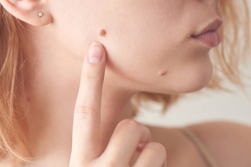
Unusual moles on the body—such as raised moles, those with hair, bleeding, blue or red in color, or unusually large in size—can often raise concerns about skin cancer. Dermatologist Dr. La Duong from Quoc Thai General Hospital (Taiwan), via Sohu, has identified common types of moles, their causes, and how to recognize signs that a mole may become cancerous.
Three Basic Types of Moles in Medicine
According to Dr. La Duong, moles originate from melanocytes and can be classified into three main types based on their location in the skin:
-
Junctional Nevus:
Found at the junction between the epidermis and dermis. These moles are usually flat and dark in color. This is the most common type of mole. -
Compound Nevus:
Located slightly deeper in the dermis, these moles are slightly raised and may grow hair. -
Intradermal Nevus:
Completely embedded in the dermis, these moles are raised, light brown or flesh-colored, often hairy, and also referred to as “flesh moles.”
Causes of Mole Formation
Moles appear due to abnormal melanocyte development. While some are congenital, they can also form due to UV exposure, hormonal drugs, immunosuppressants, or hormonal fluctuations during pregnancy and puberty.
Types of Moles That Need Special Attention
-
Blue or Gray Moles:
Often benign blue nevi, but if they appear suddenly or change shape, testing is needed to rule out melanoma. -
Congenital Moles:
These carry a risk of turning malignant.-
Moles <1.5 cm: typically low risk.
-
Moles 1.5–20 cm: require regular monitoring.
-
Moles >20 cm: should be considered for surgical removal.
-
-
Moles with a White Halo:
Could be “halo nevi” or a sign of vitiligo, where immune cells attack melanocytes. Often benign but should be monitored. -
Disappearing Moles:
May indicate the immune system is destroying melanocytes. If accompanied by other unusual symptoms, see a doctor. -
Red Moles:
Small red dots that don’t change are often cherry angiomas—benign and age-related. However, if the mole changes color or shape, get a medical checkup. -
Scratched or Bleeding Moles:
A red flag. Moles that frequently peel, bleed, itch, or don’t heal may be signs of skin cancer. -
Raised or Hairy Moles – Are They Cancerous?
Many believe hairy or raised moles are dangerous. This is not necessarily true. Moles deep in the dermis often grow hair due to proximity to hair follicles—this is normal and not directly linked to cancer. -
Can Moles Become Skin Cancer?
Dr. La Duong explains that aside from large congenital moles, most moles do not become cancerous. Most skin cancers develop as malignant skin lesions from the beginning but are often mistaken for moles because of their similar appearance.
How to Spot Skin Cancer: The ABCDE Rule
To detect early signs of skin cancer, dermatologists recommend using the ABCDE rule:
-
A – Asymmetry:
One half of the mole does not match the other. -
B – Border:
Edges are irregular, blurry, or jagged. -
C – Color:
Uneven coloring or multiple shades in a single mole. -
D – Diameter:
Larger than 6 mm or increasing rapidly in size. -
E – Evolving:
Noticeable changes in shape, color, or behavior (such as itching, bleeding, or crusting).
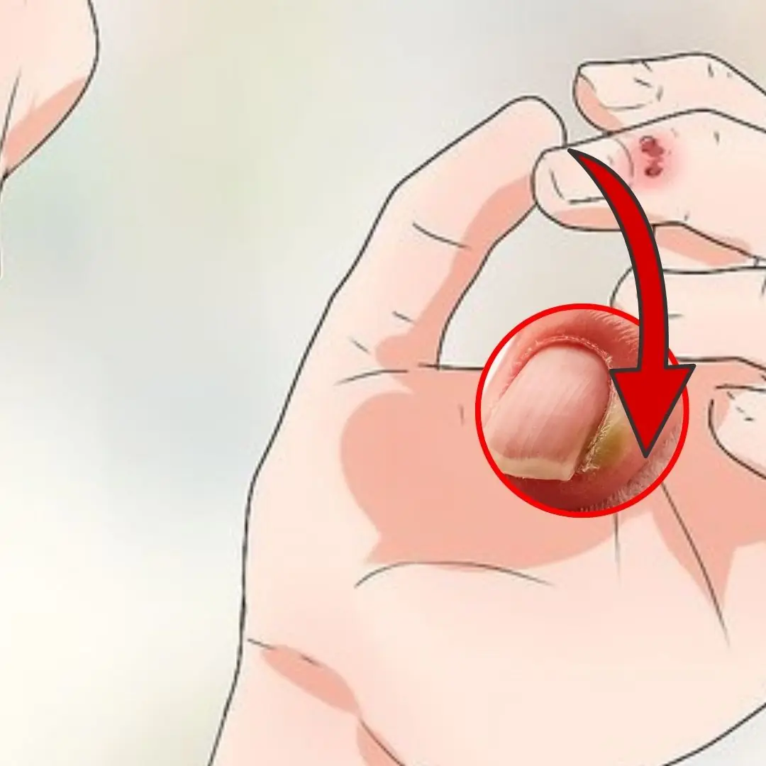
Warning: Moles on Hands and Feet Require Extra Attention
Melanoma—the most serious type of skin cancer—often appears on extremities such as fingers and toes. Therefore, be particularly cautious if moles in these areas show any unusual changes.
Expert Advice
If you notice any mole with abnormal signs based on the ABCDE rule, see a dermatologist as soon as possible. Early detection is key to effective treatment and can help prevent the risk of skin cancer in the future.